Spine MRI Quick Reference Guide for Patients
Cervical Spine MRI
MRI of the cervical spine is an extremely useful tool in evaluating patients with neck pain or symptoms of a pinched nerve as well as possible compression of the spinal cord. MRI can accurately assess for degenerative disc disease as well as disc herniation. The spinal cord itself will also be assessed for any abnormality. A specialized coil will be placed around your patient’s neck.
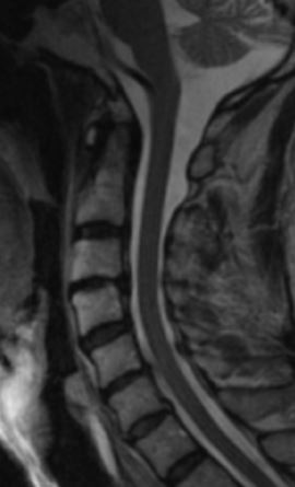
Cervical Spine MRI done by Guilford Radiology, 2010
Thoracic Spine MRI
MRI of the thoracic spine is a useful tool in evaluating patients with mid back pain or symptoms of possible compression of the spinal cord. MRI can accurately assess for degenerative disc disease as well as disc herniation. The spinal cord itself will also be assessed for any abnormality.
Lumbar Spine MRI
MRI of the lumbar spine is an extremely useful tool in evaluating patients with low back pain or symptoms of a pinched nerve. MRI can accurately assess for degenerative disc disease as well as disc herniation.
MRI of the spine looks at the vertebrae that make up the spine, as well as the disks, spinal cord, and the spaces between the vertebrae through which the nerves pass.
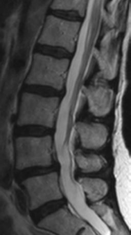
MRI Lumbar Spine, done by Guilford Radiology, 2010
Patient Preparation
For the MRI exam, if claustrophobia or anxiety is a problem, your referring physician may wish to prescribe a mild sedative to be given prior to the study. No other pre-visit preparation is necessary. You will need to remove all jewelry, hairclips, pony-tails and bobby pins. In addition, you should avoid wearing any clothes with metal. You will need to remove all clothing containing metal. This would include bras with metal enclosures and jeans with metal zippers and buttons. You will be provided a gown and a secure locker in which valuables can be placed.
CPT Codes:
- 72148 L-Spine Without Contrast
- 72158 L-Spine Without and With Contrast
- 72141 C-Spine Without Contrast
- 72156 C-Spine Without and With Contrast
- 72146 T-Spine Without Contrast
- 72157 T-Spine Without and With Contrast
Patient Weight Limit
Our MRI equipment has a weight limit of 440 pounds.
Did your doctor order this exam?
General Information
MR imaging uses a powerful magnetic field, radio frequency pulses and a computer to produce detailed pictures of organs, soft tissues, bone and virtually all other internal body structures. MRI does not use ionizing radiation (x-rays). Detailed MR images allow physicians to better evaluate various parts of the body and determine the presence of certain diseases that may not be assessed adequately with other imaging methods.
Exam Indications
Without Contrast: neck pain, mid-back pain, numbness or tingling of the arms or fingers, pain
With and Without Contrast: history of MS, transverse myelitis, tumors, cancer, post operative
Contraindications
Patients with cardiac pacemakers, ICD, or neuro-stimulators CAN NOT have an MRI. Patients with pins, plates, screws and joint replacements, stents & filters can have an MRI as long as it has been 6 weeks since placement of the device. Women who are pregnant should avoid having an elective MRI. Women who are pregnant and need an MRI should be individually evaluated for risk vs. benefits and should avoid an MRI in the 1st trimester of pregnancy.
Risks
Although the strong magnetic field is not harmful in itself, implanted medical devices that contain metal may malfunction or cause problems during an MRI exam.
There is a very slight risk of an allergic reaction if contrast material is injected. Such reactions usually are mild and easily controlled by medication. If you experience allergic symptoms, a radiologist or other physician will be available for immediate assistance.
Nephrogenic systemic fibrosis is currently a recognized, but rare, complication of MRI believed to be caused by the injection of high doses of MRI contrast material in patients with very poor kidney function.
What Happens During the Test?
You will be asked to lie down on his back on the scanning table. The table will then slide into the scanning area. During the test, the MRI will make a rapid tapping noise. Some MRI examinations may require an injection of contrast material into a vein in the arm. Your experience and comfort are of key importance. Therefore, you can watch TV, be offered earplugs or a music headset; in addition blankets are also available. You should relax and remain still during the exam. Plan 60-90 minutes of total clinic time. The scan time can vary from 30-60 minutes depending on the study. You may resume normal activities following the MRI.
The Results
A radiologist will analyze the images and send a signed report to the referring physician within 1 business day.
Questions?
If you have any questions or concerns about your procedure, feel free to call us at 203-453-5123 or contact us online.