Thyroid Ultrasound Quick Reference Guide for Patients
Ordering Information:
An ultrasound of the thyroid is typically used to help evaluate:
- enlarged thyroid
- palpable mass
- abnormal thyroid enzymes
- abnormalities seen on other modalities
- dysphasia
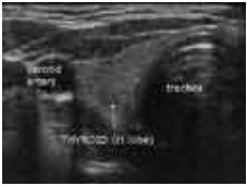
Thyroid nodules are common and occur in up to 50% of the adult population; however, less than 7% of thyroid nodules are malignant. High-resolution ultrasonography (US) is commonly used to evaluate the thyroid gland and to determine if there is a focal mass in the thyroid, or there is diffuse enlargement present. It can be used to determine if a lump is solid or a cyst. Sometimes, a fine needle biopsy will be needed to determine if a solid lump is cancer, or possible a follow-up scan.
CPT Code:
There is only one CPT code for all thyroid ultrasounds: 76536
General Information
What is Ultrasound?
Ultrasound imaging or sonography, involves exposing part of the body to high-frequency sound waves to produce pictures of the inside of the body. Ultrasound exams do not use x-rays. Because ultrasound images are captured in real-time, they can show the structure and movement of the body's internal organs, as well as blood flowing through blood vessels.
Ultrasound imaging is a noninvasive medical test that helps physicians diagnose and treat medical conditions.
Doppler ultrasound is a special ultrasound technique that evaluates blood flow through a blood vessel, including the body's major arteries and veins in the abdomen, arms, legs and neck.
Ultrasound scanners consist of a console containing a computer and electronics, a video display screen and a transducer that is used to scan the body and blood vessels. The transducer is a small hand-held device that resembles a microphone, attached to the scanner by a cord. The transducer sends out high frequency sound waves into the body and then listens for the returning echoes from the tissues in the body. The principles are similar to sonar used by boats and submarines.
The ultrasound image is immediately visible on a nearby video display screen that looks much like a computer or television monitor. The image is created based on the amplitude (strength), frequency and time it takes for the sound signal to return from the patient to the transducer and the type of body structure the sound travels through.
How does the procedure work?
Ultrasound imaging is based on the same principles involved in the sonar used by bats, ships and fishermen. When a sound wave strikes an object, it bounces back, or echoes. By measuring these echo waves it is possible to determine how far away the object is and its size, shape, and consistency (whether the object is solid, filled with fluid, or both).
In medicine, ultrasound is used to detect changes in appearance of organs, tissues, and vessels or detect abnormal masses, such as tumors.
In an ultrasound examination, a transducer both sends the sound waves and records the echoing waves. When the transducer is pressed against the skin, it directs small pulses of inaudible, high-frequency sound waves into the body. As the sound waves bounce off of internal organs, fluids and tissues, the sensitive microphone in the transducer records tiny changes in the sound's pitch and direction. These signature waves are instantly measured and displayed by a computer, which in turn creates a real-time picture on the monitor. One or more frames of the moving pictures are typically captured as still images. Doppler ultrasound, a special application of ultrasound, measures the direction and speed of blood cells as they move through vessels. The movement of blood cells causes a change in pitch of the reflected sound waves (called the Doppler effect). A computer collects and processes the sounds and creates graphs or color pictures that represent the flow of blood through the blood vessels.
How is the procedure performed?
For most ultrasound exams, you are positioned lying face-up on an examination table that can be tilted or moved.
A clear water-based gel is applied to the area of the body being studied to help the transducer make secure contact with the body and eliminate air pockets between the transducer and the skin. The sonographer (ultrasound technologist) then presses the transducer firmly against the skin in various locations, sweeping over the area of interest or angling the sound beam from a farther location to better see an area of concern.
Doppler sonography is performed using the same transducer.
When the examination is complete, you may be asked to dress and wait while the ultrasound images are reviewed. This ultrasound examination is usually completed within 30 to 60 minutes.
Questions?
If you have any questions or concerns about your procedure, feel free to call us at 203-453-5123 or contact us online.