CT Extremities Cat Scan Quick Reference Guide for Patients
Upper & Lower Extremities
Extremities are the fancy medical term for our arms and legs. CT of the extremities is excellent for examining bone detail and anatomy, particularly in the evaluation of fractures, osteomyelitis, and bone tumors, as well as for arthritis and chronic pain. It is also an excellent alternative to MRI for the evaluation of the soft tissues, in patients who can’t have an MRI exam.
These studies are often done without contrast, except for osteomyelitis and tumor, which are done without and with contrast. For joint evaluation, consider a CT arthrogram, described under a separate Physician Quick Reference Guide.
Upper Extremities CT examines the hand, wrist, forearm, elbow, humerus, shoulder and clavicle. Lower Extremities CT examines the hip, knee, ankle and foot.
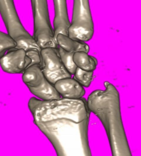
A 3D wrist CT performed at Guilford Radiology, 2010
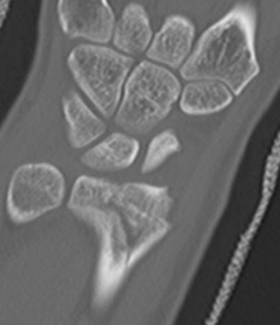
A wrist CT performed at Guilford Radiology, 2010
Patient Preparation
Non Contrast: no preparation
Intravenous Contrast Exams:
You will be instructed to not eat solid foods for 4 hours prior to the intravenous contrast injection.
A creatinine (within 6 months) is required if the patient is over 50 years of age or older.
Patients on metformin and other oral diabetic medication must not be taken for 48 hours after the CT. Your patient may need a blood test (creatinine level) to check renal functions prior to restarting the medication. The CT Technologist will contact the referring physician at the time of the examination to review follow-up instructions.
CPT Codes
Upper Extremities CT:
- 73200 Without Contrast
- 73201 With Contrast
- 73202 With and Without Contrast
Lower Extremities CT:
- 73700 Without Contrast
- 73701 With Contrast
- 73702 Without and With Contrast
Patient Weight Limit
Our CT Scan tables have a weight limit of 660 pounds.
Did your doctor order this exam?
General Information about CT Scanning
What is CT scanning?
CT scanning combines special x-ray equipment with sophisticated computers to produce multiple images of the inside of the body. These cross-sectional images are then examined on a computer monitor by a radiologist. They also can be printed or transferred to a CD. CT scans of internal organs, bones, soft tissue and blood vessels provide greater clarity and reveal more details than regular x-ray exams. Using specialized equipment and expertise to create and interpret CT scans of the body, radiologists can more easily diagnose problems such as cancers, cardiovascular disease, infectious disease, appendicitis, trauma and musculoskeletal disorders.
There has been considerable work done recently on radiation dose from CT scans. At our offices, our CT scanners adjust the radiation dose for each patient to use the lowest possible dose.
How does the procedure work?
In many ways CT scanning works very much like other x-ray examinations. Different body parts absorb the x-rays in varying degrees.
In a conventional x-ray exam, a small burst of radiation is aimed at and passes through the body, recording an image on photographic film or a special image recording plate. Bones appear white on the xray; soft tissue shows up in shades of gray and air appears black.
With CT scanning, numerous x-ray beams and a set of electronic x-ray detectors rotate around the patient, measuring the amount of radiation being absorbed throughout his/her body. At the same time, the examination table is moving through the scanner, so that the x-ray beam follows a spiral path. A special computer program processes this large volume of data to create two-dimensional cross-sectional images of the body, which are then displayed on a monitor. This technique is called helical or spiral CT.
The CT scanner at our office is a 80 slice multidetector scanner, allowing thinner slices to be obtained in a shorter period of time, resulting in more detail and additional view capabilities. Our scanner is so fast it can scan through large sections of the body in just a few seconds. Such speed is beneficial for all patients but especially children, the elderly and critically ill. For children, the CT scanner technique will be adjusted to reduce the radiation dose. For some CT exams, a contrast material is used to enhance visibility in the area of the body being studied.
Questions?
If you have any questions or concerns about your procedure, feel free to call us at 203-453-5123 or contact us online.