CT Arthrography Cat Scan Arthrogram Quick Reference Guide for Physicians
CT arthrography can be used as an alternative to MRI arthrography in patients who cannot have an MRI exam. This exam involves the injection of a contrast material into the joint. An CT exam of the joint is performed immediately after the contrast injection, which shows the fine details of the joint space. Common indications for CT arthrography include the post-operative knee and shoulder, evaluation of the shoulder and hip labra, and for the evaluation of the ligaments of the wrist. The contrast solution injected into the patient contains a small amount of iodinated.
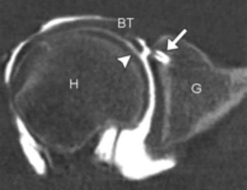
Image from a CT Arthrogram study done by Guilford Radiology, 2010
Patient Preparation
No preparation is required.
CPT Codes
- 70450 Without Contrast
- 70470 Without and With Contrast
Patient Weight Limit
Our CT Scan tables have a weight limit of 660 pounds.
Ready to Order a Test for your Patient?
- Download Requisition Form Here (Please write CT Arthrogram in the “other” section and indicate joint to be scanned.)
General Information about CT Scanning
What is CT scanning?
CT scanning combines special x-ray equipment with sophisticated computers to produce multiple images of the inside of the body. These cross-sectional images are then examined on a computer monitor by a radiologist. They also can be printed or transferred to a CD. CT scans of internal organs, bones, soft tissue and blood vessels provide greater clarity and reveal more details than regular x-ray exams. Using specialized equipment and expertise to create and interpret CT scans of the body, radiologists can more easily diagnose problems such as cancers, cardiovascular disease, infectious disease, appendicitis, trauma and musculoskeletal disorders.
There has been considerable work done recently on radiation dose from CT scans. At our offices, our CT scanners adjust the radiation dose for each patient to use the lowest possible dose.
How does the procedure work?
In many ways CT scanning works very much like other x-ray examinations. Different body parts absorb the x-rays in varying degrees.
In a conventional x-ray exam, a small burst of radiation is aimed at and passes through the body, recording an image on photographic film or a special image recording plate. Bones appear white on the xray; soft tissue shows up in shades of gray and air appears black.
With CT scanning, numerous x-ray beams and a set of electronic x-ray detectors rotate around the patient, measuring the amount of radiation being absorbed throughout his/her body. At the same time, the examination table is moving through the scanner, so that the x-ray beam follows a spiral path. A special computer program processes this large volume of data to create two-dimensional cross-sectional images of the body, which are then displayed on a monitor. This technique is called helical or spiral CT.
The CT scanners at our offices are multidetector scanners, allowing thinner slices to be obtained in a shorter period of time, resulting in more detail and additional view capabilities. Our scanners are so fast that they can scan through large sections of the body in just a few seconds. Such speed is beneficial for all patients but especially children, the elderly and critically ill. For children, the CT scanner technique will be adjusted to reduce the radiation dose. For some CT exams, a contrast material is used to enhance visibility in the area of the body being studied.
What Happens During the CT Arthrography?
For the contrast injection, the radiologist will explain the procedure to the patient, and obtain consent for the procedure. The patient is positioned on the x-ray examination table in the fluoroscopy room. Next, the skin around the joint is cleansed with antiseptic and covered with a sterile drape. The skin and soft tissues are numbed by a local anesthetic injected into the area. A needle is then inserted through this numbed skin into the joint space. Contrast material is injected into the joint space and the needle is removed. The patient will experience a slight pinprick and may feel a momentary burning from the local anesthesia used to numb the area. The patient may feel a fullness as the joint is filled with contrast.
For the actual CT arthrogram study, the patient is then transferred to the CT scanner. Your patient will be asked to lie down on his back on the scanning table. The table will then slide into the scanning area. Your patient’s experience and comfort are of key importance. The scan time can vary from 30-60 minutes depending on the study. Your patient may resume normal activities following the CT.
Women should always inform their physician and x-ray technologist if there is any possibility that they are pregnant. Many imaging tests are not performed during pregnancy so as not to expose the fetus to radiation.