


Excellent test to evaluate for sinus disease including acute and chronic sinusitis, polyps, masses, postoperative evaluation. Usually done without contrast. Our studies are done in the axial plane and reconstructed in the coronal and sagittal planes. 3-D reconstructions are done as needed.
No preparation required.
70486 Without Contrast
Our CT Scan tables have a weight limit of 660 pounds.
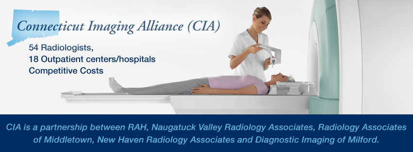
CT scanning combines special x-ray equipment with sophisticated computers to produce multiple images of the inside of the body. These cross-sectional images are then examined on a computer monitor by a radiologist. They also can be printed or transferred to a CD. CT scans of internal organs, bones, soft tissue and blood vessels provide greater clarity and reveal more details than regular x-ray exams. Using specialized equipment and expertise to create and interpret CT scans of the body, radiologists can more easily diagnose problems such as cancers, cardiovascular disease, infectious disease, appendicitis, trauma and musculoskeletal disorders.
There has been considerable work done recently on radiation dose from CT scans. At our offices, our CT scanners adjust the radiation dose for each patient to use the lowest possible dose.
In many ways CT scanning works very much like other x-ray examinations. Different body parts absorb the x-rays in varying degrees.
In a conventional x-ray exam, a small burst of radiation is aimed at and passes through the body, recording an image on photographic film or a special image recording plate. Bones appear white on the xray; soft tissue shows up in shades of gray and air appears black.
With CT scanning, numerous x-ray beams and a set of electronic x-ray detectors rotate around the patient, measuring the amount of radiation being absorbed throughout his/her body. At the same time, the examination table is moving through the scanner, so that the x-ray beam follows a spiral path. A special computer program processes this large volume of data to create two-dimensional cross-sectional images of the body, which are then displayed on a monitor. This technique is called helical or spiral CT.
The CT scanner at our office is a 80 slice multidetector scanner, allowing thinner slices to be obtained in a shorter period of time, resulting in more detail and additional view capabilities. Our scanner is so fast it can scan through large sections of the body in just a few seconds. Such speed is beneficial for all patients but especially children, the elderly and critically ill. For children, the CT scanner technique will be adjusted to reduce the radiation dose. For some CT exams, a contrast material is used to enhance visibility in the area of the body being studied.
Phone:
(203) 453-5123
Hours:
M-F 7:30AM to 5PM
Sat. 8AM to Noon