Carotid Ultrasound Quick Reference Guide for Physicians
The carotid ultrasound is most frequently performed to detect narrowing, or stenosis, of the carotid artery, a condition that substantially increases the risk of stroke. The major goal of carotid ultrasound is to screen patients for blockage or narrowing of their carotid arteries, which if present may increase their risk of having a stroke. Once the diagnosis is made a comprehensive treatment may be initiated. It may also be performed if a patient has high blood pressure or a carotid bruit—an abnormal sound in the neck that is heard with the stethoscope. Other risk factors calling for a carotid ultrasound are:
- Advanced age
- Diabetes
- Elevated blood cholesterol
- A family history of stroke or heart disease
A carotid ultrasound is performed to evaluate for the following:
- Narrowing of vessels (which may be caused by plaque).
- Blockages to blood flow (such as clots).
- Locating a hematoma, a collection of clotted blood that may slow and eventually stop blood flow.
- Dissection of the carotid artery, a split between layers of the artery wall that may lead to obstruction of blood flow or a weakening of the wall of the artery.
- To check the state of the carotid artery after surgery to restore normal blood flow.
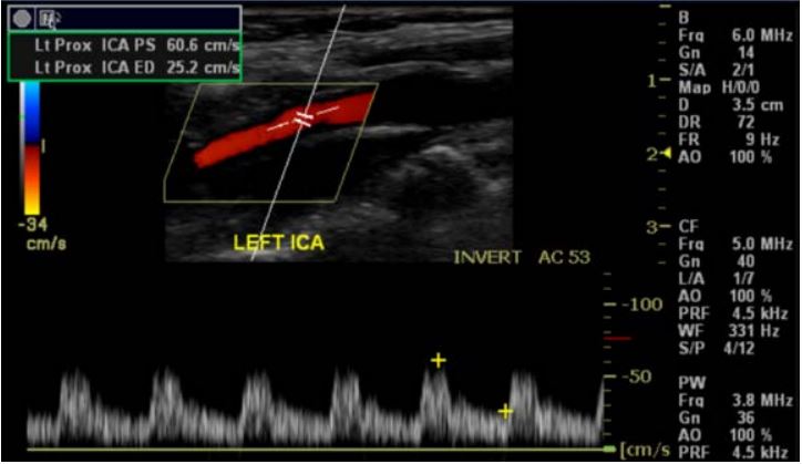 Image from a Carotid Ultrasound done at Guilford Radiology, 2009 No preparation by your patient is required.
There are no contraindications for Carotid Ultrasound.
CPT codes: 93880 bilateral
93882 unilateral
Questions? Please call Guilford Radiology at (860)453-5123 or West Haven Radiology at (860)934-4482 if you would like to speak with the on-site Radiologist, a technologist for the specific modality in which you are interested, or another member of our team. Or, click here for information on contacting our Physician Liaison Team, who will promptly respond to your questions.
Ready to Order a Test for your Patient?
General Information
What is Ultrasound?
Ultrasound imaging or sonography, involves exposing part of the body to high-frequency sound waves to produce pictures of the inside of the body. Ultrasound exams do not use x-rays. Because ultrasound images are captured in real-time, they can show the structure and movement of the body's internal organs, as well as blood flowing through blood vessels. Ultrasound imaging is a noninvasive medical test that helps physicians diagnose and treat medical conditions.
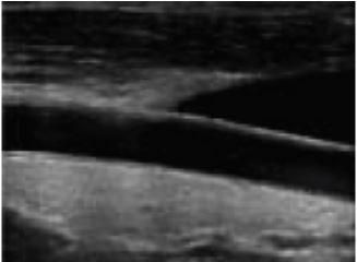
An ultrasound view of the carotid artery
Doppler ultrasound is a special ultrasound technique that evaluates blood flow through a blood vessel, including the body's major arteries and veins in the abdomen, arms, legs and neck.
Ultrasound scanners consist of a console containing a computer and electronics, a video display screen and a transducer that is used to scan the body and blood vessels. The transducer is a small hand-held device that resembles a microphone, attached to the scanner by a cord. The transducer sends out high frequency sound waves into the body and then listens for the returning echoes from the tissues in the body. The principles are similar to sonar used by boats and submarines.
The ultrasound image is immediately visible on a nearby video display screen that looks much like a computer or television monitor. The image is created based on the amplitude (strength), frequency and time it takes for the sound signal to return from the patient to the transducer and the type of body structure the sound travels through.
How does the procedure work?
Ultrasound imaging is based on the same principles involved in the sonar used by bats, ships and fishermen. When a sound wave strikes an object, it bounces back, or echoes. By measuring these echo waves it is possible to determine how far away the object is and its size, shape, and consistency (whether the object is solid, filled with fluid, or both).
In medicine, ultrasound is used to detect changes in appearance of organs, tissues, and vessels or detect abnormal masses, such as tumors.
In an ultrasound examination, a transducer both sends the sound waves and records the echoing waves. When the transducer is pressed against the skin, it directs small pulses of inaudible, high-frequency sound waves into the body. As the sound waves bounce off of internal organs, fluids and tissues, the sensitive microphone in the transducer records tiny changes in the sound's pitch and direction. These signature waves are instantly measured and displayed by a computer, which in turn creates a real-time picture on the monitor. One or more frames of the moving pictures are typically captured as still images. Doppler ultrasound, a special application of ultrasound, measures the direction and speed of blood cells as they move through vessels. The movement of blood cells causes a change in pitch of the reflected sound waves (called the Doppler effect). A computer collects and processes the sounds and creates graphs or color pictures that represent the flow of blood through the blood vessels.
How is the procedure performed?
For most ultrasound exams, the patient is positioned lying face-up on an examination table that can be tilted or moved.
A clear water-based gel is applied to the area of the body being studied to help the transducer make secure contact with the body and eliminate air pockets between the transducer and the skin. The sonographer (ultrasound technologist) then presses the transducer firmly against the skin in various locations, sweeping over the area of interest or angling the sound beam from a farther location to better see an area of concern. Doppler sonography is performed using the same transducer.
When the examination is complete, the patient may be asked to dress and wait while the ultrasound images are reviewed. This ultrasound examination is usually completed within 30 to 60 minutes.
Information adapted from www.radiologyinfo.org, reviewed and edited by Michael Crain, MD.
This manual is intended for use as merely a guideline for referring physicians and their staff only. It contains information pertaining to the most commonly ordered exams and indications. RAM Radiology does not recommend any particular examination. Individual radiologist preference or patient medical information may dictate ordering alternative studies.