Abdomen MRI Quick Reference Guide for Physicians
MRI of the abdomen generally is used to evaluate individual organs, and it is an extremely important tool for radiologists. Most MRI examinations of the abdomen are organ specific, ie, liver, kidney, adrenal gland, and are ordered without and with contrast, when possible. CT scanning is more useful in the workup of nonspecific regional complaints, such as "upper abdominal pain" that is not necessarily associated with one organ.
MRI's superior soft tissue contrast makes it excellent at detecting and characterizing diseases of the liver. MRI is able to distinguish between benign masses (such as hemangiomas or adenomas) and malignancies. MRI can do this with a high degree of accuracy, and frequently eliminate the need for liver biopsy.
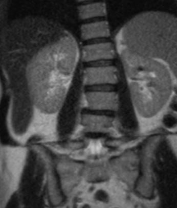
In the kidneys, MRI is frequently used to distinguish between benign lesions (such as cysts) and malignancies. Because fluid and fluid containing lesions such as cysts are common lesions in kidneys, it is important to distinguish them from potential cancers, and MRI can do this quite well.
MRI is exceptionally suited to differentiate between benign adrenal masses, which are common and called adenomas, and malignancies, which spread frequently to the adrenal glands. With a high degree of accuracy, MRI can tell whether the adrenal glands are involved in the spread of cancer, or contain merely a benign adrenal adenoma.
When an MRI of the abdomen is ordered, the organ to be visualized should be specified.
MRI of the Abdomen, done at Guilford Radiology, 2010
CPT Codes:
- 74181 Without Contrast
- 74183 Without and With Contrast
Patient Weight Limit
Our MRI equipment has a weight limit of 440 pounds.
Ready to Order a Test for your Patient?
General Information
MR imaging uses a powerful magnetic field, radio frequency pulses and a computer to produce detailed pictures of organs, soft tissues, bone and virtually all other internal body structures. MRI does not use ionizing radiation (x-rays). Detailed MR images allow physicians to better evaluate various parts of the body and determine the presence of certain diseases that may not be assessed adequately with other imaging methods.
Contraindications
Patients with cardiac pacemakers, ICD, or neuro-stimulators CAN NOT have an MRI. Patients with pins, plates, screws and joint replacements, stents & filters can have an MRI as long as it has been 6 weeks since placement of the device. Women who are pregnant should avoid having an elective MRI. Women who are pregnant and need an MRI should be individually evaluated for risk vs. benefits and should avoid an MRI in the 1st trimester of pregnancy.
How Does Your Patient Prepare?
A current creatinine (within 45 days) is needed on all patients over the age of 60, as well as patients that have high blood pressure, diabetes, acute vascular disease, or a history of kidney disease. Please fax these results with the order. Patients will need to remove all jewelry, hairclips, pony-tails and bobby pins. In addition, the patient will need to remove all clothing containing metal. This would include bras with metal enclosures and jeans with metal zippers and buttons. Your patient will be provided a gown and a secure locker in which valuables can be placed.
Risks
Although the strong magnetic field is not harmful in itself, implanted medical devices that contain metal may malfunction or cause problems during an MRI exam.
There is a very slight risk of an allergic reaction if contrast material is injected. Such reactions usually are mild and easily controlled by medication. If you experience allergic symptoms, a radiologist or other physician will be available for immediate assistance.
Nephrogenic systemic fibrosis is currently a recognized, but rare, complication of MRI believed to be caused by the injection of high doses of MRI contrast material in patients with very poor kidney function.
What Happens During the Test?
Your patient will be asked to lie down on his back on the scanning table. The table will then slide into the scanning area. During the test, the MRI will make a rapid tapping noise. Some MRI examinations may require an injection of contrast material into a vein in the arm. Your patient’s experience and comfort are of key importance. Therefore, our patients are offered earplugs or a music headset; in addition blankets are also available. Your patient should relax and remain still during the exam. Your patient should plan 60-90 minutes of total clinic time. The scan time can vary from 30-60 minutes depending on the study. Your patient may resume normal activities following the MRI.
The Results
A radiologist will analyze the images and send a signed report to the referring physician within 1 business day.